Invited Talks Abstracts & Key words
- Session I. 10:00 ~ 12:00
- Short Talk Session 13:30 ~ 13:45
- Session II. 13:45 ~ 15:30
- Session III. 15:50 ~ 17:50
Session I.
Chair: Miki Ebisuya (CDB, RIKEN,Japan)
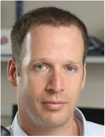 |
Dr. Avigdor Eldar |
| Tel Aviv University, Israel | |
| Talk Title: Social evolution shapes the diversification of bacterial intercellular signaling | |
| Author: Avigdor Eldar | |
| Abstract: | Microbial ‘quorum sensing’ (QS) systems, where microbes produce and respond to a signaling molecule, enable cells to sense their local density and coordinate a cooperative response to their environment.
QS-dependent cooperation is prone to exploitation by a signal reception mutant that avoids the costs of Quorum-response but rip the cooperative benefits.
Cheater null alleles are rare in wild isolates but in contrast, many QS systems show intraspecific allelic divergence of receptor-signal locus in which all alleles are functional but a signaling molecule from one allelic type (known as pherotype) activates its cognate receptor but fails to activate those of other receptor variants in the same species.
The phylogeny of the QS locus often deviates from the house-keeping phylogeny, indicating some level of selection for horizontal gene transfer (HGT).
It is unclear what evolutionary mechanisms explain the lack of null alleles and the existence and phylogenetic patterns of multiple different pherotypes and how does this evolutionary pattern relate to the cooperative behavior of the cells.
In this talk I would present a combination of bioinformatical analysis, theoretical modeling and experiments in the model bacteria /B. subtilis/ that explain this evolutionary pattern.
We demonstrate that a /B. subtilis/ reception null mutant is a cheater of the wild-type – in well-mixed environments, it invades into the wild-type population and lead to a general reduction of fitness.
The wild-type resist the invasion if the population is spatially structured.
When strains from different pherotypes compete, we find that the minority pherotype always invade the majority pherotype and that this invasion will occur also in highly structured environment.
Our results can be explained by a model that requires a QS-dependent cooperative behavior.
We therefore conclude that QS is a cooperative trait but that the strong structure of the environment prevents the evolution of cheaters, while allowing the evolution of new pherotypes and their rapid spread through horizontal gene transfer.
|
| Keywords: |
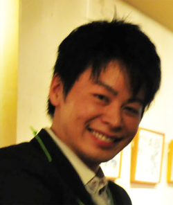 |
Dr. Naoki Irie |
| University of Tokyo, Japan | |
| Talk Title: Flexibility in early animal developmental system | |
| Author: Naoki Irie | |
| Abstract: | Recent advancements in developmental biology highlighted cascade-like molecular mechanisms taking place during embryogenesis, once again supporting our “mechanistic view of life”. On the other hand, apparent discrepancy with this viewpoint was suggested form recent studies in the field of Evo-Devo (Evolutionary Developmental Biology). Taking advantages of state of art technologies, these studies, including ours culminated in tackling the issue of classic argument on how the relationship between ontogeny and phylogeny can be formulated. Our study demonstrated that the hourglass model best explains the divergence pattern of gene expression profiles between embryos of four vertebrate species (mouse, chicken, xenopus, and zebrafish), and for the first time, identified the stages having the most conserved expression profiles, namely, pharyngular stages. This was further tested with the animal of unique body plan: turtle. Starting from genome sequencing, we will provide our current progress on this project together with discussion. Finally, the bottleneck period of the hourglass suggests that animal embryogenesis somehow tolerated evolutionary divergence in early stages, while keeping mid embryonic stages conserved. This further implies unexpected characteristics of animal embryogenesis, or highly flexible characteristics of early developmental system. |
| Keywords: | Evo-Devo, hourglass model |
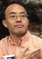 |
Dr. Yu-Chiun Wang |
| CDB, RIKEN, Japan | |
| Talk Title: Quantitative 4D analyses of epithelial folding during Drosophila gastrulation | |
| Author:
Zia Khan [1], Yu-Chiun Wang [2,3,4], Eric F. Wischaus [2,3] and Matthias Kaschube [5]
[1] Department of Human Genetics, University of Chicago, Chicago, IL 60637, USA [2] Department of Molecular Biology, Princeton University, Princeton, NJ 08544, USA [3] The Howard Hughes Medical Institute, Moffett Laboratory 435, Princeton University, Princeton, NJ 08544, USA [4] Laboratory for Epigenetic Morphogenesis, RIKEN Center for Developmental Biology, Kobe, Hyogo 650-0047, Japan [5] Frankfurt Institute for Advanced Studies, Faculty of Computer Science and Mathematics, Goethe University, D-60438 Frankfurt am Main Germany |
|
| Abstract: |
Quantitative 3D analyses of cellular dynamics using time-lapse microscopy data have the potential to
greatly advance our understanding of complex morphogenetic processes. Yet, obtaining these
measurements remains technically challenging due to the intrinsic noise in these data. We developed
a new software tool called EDGE4D that automates segmentation, tracking, and quantitative
measurement of cellular properties at single cell resolution. EDGE4D introduces several novel
algorithmic strategies, such as techniques that leverage the duality between surface meshes and
binary image volumes, that allow reliable, systematic, and dynamic analyses of a large number of
densely packed membrane-labeled cells, analyses that were previously unattainable by existing
methods. We used EDGE4D in a pilot study that tackles the complex cell shape changes and rapid
tissue movements that occur during the formation of the dorsal folds in the gastrulating Drosophila
embryo. Although the depth of the tissue structure and the poor signal-to-noise imaging conditions
render quantitative analysis challenging, EDGE4D successfully confirms known features of this
morphogenetic event and uncovers previously undocumented patterns of tissue and cellular dynamics.
Furthermore, EDGE4D enables the first 3D, dynamic analysis of cell contact surfaces, allowing us to
propose new hypotheses on how internal and external forces might contribute to cell shape changes
during dorsal fold formation. EDGE4D addresses a growing need for new generally applicable tools and
algorithms for processing and quantitation of 3D time-lapse imaging data. Tools such as EDGE4D hold
the promise to transform recent advances in time-lapse imaging technologies into powerful means of
biological discovery.
|
| Keywords: | Automated image processing, 3D reconstruction of membrane-labeled cells, Cell shape change, Epithelial folding, Drosophila gastrulation |
Short Talk Session
Co-Chairs: Noriko Hiroi (Keio University, Japna) and Akira Funahashi (Keio University, Japan)
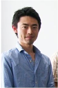 |
Masahi Kajita |
| Institute of Industrial Science, the University of Tokyo, Japan | |
| Talk Title: Modeling of self and non-self discriminaiton by T-cells. | |
| Author: Masashi Kajita, Kazuyuki Aihara and Tetsuya J. Kobayashi | |
| Abstract: | T-cells prevent us from infection or autoimmune disease by the ability to discriminate self and non-self antigens. Self and non-self discrimination is based on the interaction between antigens represented on the surface of antigens has revealed that self and non-self antigens are almost same in their structures. A parameter that characterizes the difference of antigens is the affinity between an antigen and a T-cell receptor, but this difference is very small because of the similarity in the structure. Therfore T-cell must have the ability to amplify the slight difference in affinities of antigens. There are some models for molecular discrimination that amplifies the difference of molecules exponentially, but they cannot reproduce several important phenomena. In this research, we develop a novel model that discriminates self and non-self antigens based on a different mechanism. Pur model reproduces the phenomena that the previous models cannot reproduce. We also report the several characteristics of this model and the mechanism how the system improves the accuracy of discriminaiton.
|
| Keywords: |
Session II.
Chair: Chun-Biu Li (Hokkaido University, Japan)
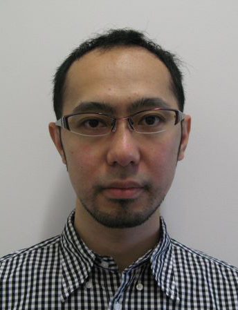 |
Dr. Yoshiyuki Arai |
| Osaka University, Japan | |
| Talk Title: Realtime fluorescence and chemiluminescence imaging with optogenetic activation in living cells. | |
| Author: Yoshiyuki Arai | |
| Abstract: | tba.
|
| Keywords: | Fluorescent probe, chemiluminescnet probe, optogenetic tool, CCD dead time |
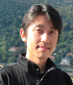 |
Dr. Hiroaki Takagi |
| Nara Medical University, Janapn | |
| Talk Title: Relevance of spontaneous migration to tactic response in Dictyostelium cells | |
| Author: Hiroaki Takagi | |
| Abstract: | We organized collaborative researchs between theory and experiment to answer the function of cellular spontaneous activity by adopting Dictyostelium discoideum. We focused on spontaneous cell migration, applied statistical analysis based on the theory of Brownian movement to cellular trajectories, and identified a generalized Langevin model that can reproduce the revealed statistical nature. Through the numerical simulations and theoretical analysis of this model, we predicted functional significance of fluctuation in cell migration dynamics. Then, we experimentally verified a essential relationship between random cell migration and directional cell migration under DCEF (electrotaxis). Now we apply corresponding analysis to apontaneous migration of immune T cell, in order to examine the generality of the view. |
| Keywords: | cell migration, anomalous diffusion, electrotaxis, Dictyostelium |
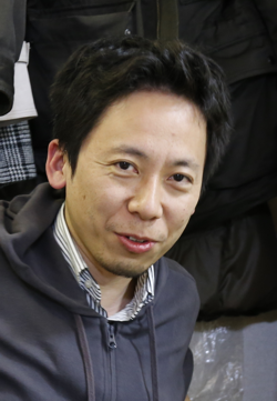 |
Dr. Rinshi Kasai |
| Institute for Frontier Medical Sciences, Kyoto University, Janapn | |
| Talk Title: Reversible dimer formation of G-protein coupled receptor: Quantitative evaluation by a single fluorescent molecule imaging | |
| Author: Rinshi Kasai | |
| Abstract: |
G-protein coupled receptor, or GPCR is one of the biggest protein families in our human body. For the past few decades, the GPCR dimerization has been discussed because of its importance regarding the signal tunings and divergences. However, there have been only few reports quantitatively showing a GPCR dimerization, resulting in ongoing controversies. Here, we wanted to resolve this controversy by applying the single fluorescent-molecule imaging technique to visualize each individual dimerization in the live plasma membrane. By directly observing GPCR dimers, their physiological properties were evaluated in terms of their dynamics. As a result of our observations of some class A GPCRs, it has eventually become clear that both monomers and dimers exist at the same time, and each dimer is transiently formed and fallen apart within a certain period, around 100 milliseconds. In this presentation, I’m going to introduce our recent progress.
|
| Keywords: |
Session III.
Chair: Ziya Kalay (iCeMS Kyoto University, Japan)
 |
Dr. Craig Jolley |
| CDB, RIKEN, Japan | |
| Talk Title: Mammalian circadian oscillations at the cellular and tissue scales | |
| Author: Craig Jolley | |
| Abstract: |
In many organisms, behavior and physiology follow an autonomous 24-hour rhythm driven by a circadian clock. In mammals, cellular oscillations are driven by transcriptional and post-translational feedback loops. In the suprachiasmatic nucleus (SCN), a specialized center in the anterior hypothalamus, the noisy oscillations of individual neurons are coupled to generate robust circadian rhythms that drive organismal behavior. Understanding SCN function is an inherently multi-scale problem: rhythms arise from molecular interactions within and between cells, but the important outputs at the level of the complete tissue, which contains ~20,000 neurons. We have employed a variety of different modeling approaches to understand both the detailed phase relationships of oscillating components within individual cells and the collective behavior of the oscillating tissue. At the single-cell level, we have developed a model in which oscillations are driven by three clock-controlled elements (CCEs) – promoter sequence elements that are bound by circadian transcription factors and give the target gene a place on the circadian schedule. Depending on which CCE sequences are present a gene will be expressed in the morning (E/E'-box), in the daytime (D-box), or in the evening (RRE); intermediate expression phases can be generated by combinatorial regulation. We used a differential evolution algorithm to identify parameter values yielding an optimal agreement with mRNA/protein expression phases derived from qPCR and quantitative Western blotting, followed by Monte Carlo sampling to determine the sensitivity of model fitting to different parameter combinations. The model outputs agree well with experimental evidence, and allow us to make novel predictions that will be tested in future experiments. At the tissue level, we are interested in how spatiotemporal “waves” of circadian gene expression arise in a heterogeneous population of SCN neurons. The SCN is known to employ a wide variety of different chemical signals, and we are building a coarse-grained model that incorporates the effects of different chemical signals. Our goal is to assess the impact of different intercellular signals on the stability, entrainability, and phase heterogeneity of the tissue-level clock. |
| Keywords: | Circadian clock, intercellular coupling, optimization, dynamical systems |
 |
Dr. Tomonobu Watanabe |
| QBiC, RIKEN, Japan | |
| Talk Title: Development of fluorescent protein to sense physical parameters | |
| Authors: Tomonobu Watanabe | |
| Abstract: |
Fluorescent protein-based indicators for intracellular environment conditions such as pH and ion concentrations are commonly used to study the status and dynamics of living cells. Despite being an important factor in many biological processes, the development of an indicator for the physicochemical state of water, such as pressure, and hydrophobicity, however, has been neglected. We here found a novel mutation that dramatically enhances the sensitivity to the state of water of the yellow fluorescent protein (YFP). In this symposium, we would like to present you the developmental strategy, proof of concept and experimental demonstrations of our fluorescent proteins.
|
| Keywords: | Fluorescent protein, pressure measurement, protein-crowding, live imaging |
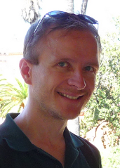 |
Dr. Andre Leier |
| OIST, Japan | |
| Talk Title: Simulating diffusion in crowded environments with multifractional Brownian motion | |
| Authors: Andre Leier | |
| Abstract: |
Diffusion of molecules within cells has many times been measured and characterized as highly anomalous. Even though the underlying mechanisms are not entirely understood, excluded volume effects inside the crowded cell have already been identified as one cause for subdiffusion. Aside from experimental studies, computer simulations have been used to deepen our understanding of anomalous diffusion. In the past, the focus has been largely on continuous time random walks (CTRWs), but recent work has suggested that fractional Brownian motion (FBM) may be a better descriptor of diffusion in crowded environments. FBM is driven by a Gaussian process with zero-mean and a covariance function that depends on the so-called Hurst exponent H. A natural generalization of FBM is achieved by replacing H by a time-dependent Hölder function H(t) leading to multifractional Brownian motion (MFBM). Here, we present results from a recent study [1] using FBM and MFBM to simulate diffusion of a tracked particle in the presence of crowding molecules and physical obstacles. Particle tracking data, mimicking experimental data, was first generated with an off-lattice particle simulator. We then attempted to obtain computationally significantly less expensive (M)FBM paths that match the statistical properties given by mean-square displacement (MSD) and time averaged MSD of our sample data. While diffusion around immovable obstacles can be reasonably characterized by a single Hurst exponent, diffusion in the presence of crowding molecules seems to exhibit multifractional properties in form of a different short and long-time behaviour. [1] T.T. Marquez-Lago, A. Leier, K. Burrage (2012). Anomalous diffusion and multifractional Brownian motion: simulating molecular crowding and physical obstacles in systems biology. IET Syst Biol (in press). |
| Keywords: | anomalous diffusion, crowding, random walks, fractional Brownian motion, spatial-stochastic modelling |
News
- 2013-09-11: IWQB2013 Abstract Booklet is now available from here. [Download]
- 2013-09-11: IWQB2013 Flyer is now available from here. [Download]
- 2013-09-13: Registration system will be available until the day before the workshop from here.
- 2013-09-11: IWQB2013 Poster is available from here. [Download]
Contact information
qbioint _at_ gmail.com
Venue
2-2 Yamadaoka, Suita
Osaka, 565-0871 JAPAN